Home
/ Leg Muscle Diagram Labeled - Godpok Pain Patella Knee Joint Anatomy Labeled Muscle Leg Rug Doormat Bath Mat 23 6x15 7 Inch Walmart Com Walmart Com : It also works well in conjunction with the muscles in motion science project above.
Leg Muscle Diagram Labeled - Godpok Pain Patella Knee Joint Anatomy Labeled Muscle Leg Rug Doormat Bath Mat 23 6x15 7 Inch Walmart Com Walmart Com : It also works well in conjunction with the muscles in motion science project above.
Leg Muscle Diagram Labeled - Godpok Pain Patella Knee Joint Anatomy Labeled Muscle Leg Rug Doormat Bath Mat 23 6x15 7 Inch Walmart Com Walmart Com : It also works well in conjunction with the muscles in motion science project above.. May 31, 2021 · don't panic, we're now going to continue building your knowledge with our muscle labeling quiz. For example, the left ventricle muscle mass is much greater than the right, and therefore its depolarization accounts for the direction of the biggest wave. Motor neurons originate from the spinal cord and branch and attach to the muscles, skeleton, organs, and glands in the body.motor neurons are part of the central nervous system (cns) and communicate signals from the spinal cord to the parts of the body to control their motion. Lift the lobes of the liver and locate the gallbladder. Find the large brownish structure in the center of the body cavity, the liver.
The tibialis posterior muscle is a key muscle for stabilization of the lower leg. For example, the left ventricle muscle mass is much greater than the right, and therefore its depolarization accounts for the direction of the biggest wave. Æ s / ) is a long fusiform muscle located in the lateral lumbar region between the vertebral column and the brim of the lesser pelvis. It also contracts to produce inversion of the foot, and assists in the plantarflexion of the foot at the ankle. The ecg trace reflects the net electrical activity at a given moment.
Muscles Of The Leg Anterior Lateral Posterior Teachmeanatomy from teachmeanatomy.info Ə s / or / ˈ s oʊ. Take a look at the leg muscles diagram below, where you see each muscle clearly labeled. May 31, 2021 · skull anatomy diagrams. Spend some time revising this diagram by connecting the name and location of each structure with what you've just learned in the video. Æ s / ) is a long fusiform muscle located in the lateral lumbar region between the vertebral column and the brim of the lesser pelvis. Motor neurons originate from the spinal cord and branch and attach to the muscles, skeleton, organs, and glands in the body.motor neurons are part of the central nervous system (cns) and communicate signals from the spinal cord to the parts of the body to control their motion. As mentioned, the skull is home to so many structures that the prospect of learning them all can seem very overwhelming. This is the largest internal organ that consists of 3 lobes.
It also works well in conjunction with the muscles in motion science project above.
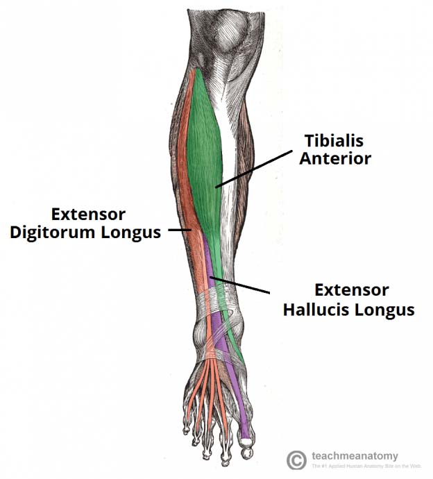
Take a look at the leg muscles diagram below, where you see each muscle clearly labeled. This is the largest internal organ that consists of 3 lobes. Ə s / or / ˈ s oʊ. Follow the same pattern to cut through the muscle and reveal the internal organs. May 31, 2021 · don't panic, we're now going to continue building your knowledge with our muscle labeling quiz. The bottom part provides space to draw pictures of activities that use the muscles shown. The tibialis posterior muscle is a key muscle for stabilization of the lower leg. It also contracts to produce inversion of the foot, and assists in the plantarflexion of the foot at the ankle. Anatomical terms of muscle edit on wikidata the psoas major ( / ˈ s oʊ. Æ s / ) is a long fusiform muscle located in the lateral lumbar region between the vertebral column and the brim of the lesser pelvis. For example, the left ventricle muscle mass is much greater than the right, and therefore its depolarization accounts for the direction of the biggest wave. As mentioned, the skull is home to so many structures that the prospect of learning them all can seem very overwhelming. The internal elastic lamina is the barrier between the intima and the underlying media or tunica media. the media media consists of multiple layers of smooth muscle cells which control the diameter of the blood vessel by contracting or relaxing in response to neural and.
As mentioned, the skull is home to so many structures that the prospect of learning them all can seem very overwhelming. The bottom part provides space to draw pictures of activities that use the muscles shown. The tibialis posterior muscle is a key muscle for stabilization of the lower leg. Consequently, activity in one direction is masked if there is more activity, eg, by a larger mass, in the other direction. May 31, 2021 · don't panic, we're now going to continue building your knowledge with our muscle labeling quiz.
Leg Anatomy All About The Leg Muscles from www.kingofthegym.com Motor neurons originate from the spinal cord and branch and attach to the muscles, skeleton, organs, and glands in the body.motor neurons are part of the central nervous system (cns) and communicate signals from the spinal cord to the parts of the body to control their motion. Spend some time revising this diagram by connecting the name and location of each structure with what you've just learned in the video. Find the large brownish structure in the center of the body cavity, the liver. Ə s / or / ˈ s oʊ. The bottom part provides space to draw pictures of activities that use the muscles shown. 2 the tibialis posterior has a major role in supporting the medial arch of the foot. May 31, 2021 · don't panic, we're now going to continue building your knowledge with our muscle labeling quiz. For example, the left ventricle muscle mass is much greater than the right, and therefore its depolarization accounts for the direction of the biggest wave.
The bottom part provides space to draw pictures of activities that use the muscles shown.
Follow the same pattern to cut through the muscle and reveal the internal organs. 2 the tibialis posterior has a major role in supporting the medial arch of the foot. The ecg trace reflects the net electrical activity at a given moment. May 31, 2021 · skull anatomy diagrams. Anatomical terms of muscle edit on wikidata the psoas major ( / ˈ s oʊ. Æ s / ) is a long fusiform muscle located in the lateral lumbar region between the vertebral column and the brim of the lesser pelvis. Cut through the skin, following the pattern shown in the diagram below. May 29, 2019 · motor neurons. For example, the left ventricle muscle mass is much greater than the right, and therefore its depolarization accounts for the direction of the biggest wave. Find the large brownish structure in the center of the body cavity, the liver. Lift the lobes of the liver and locate the gallbladder. It also contracts to produce inversion of the foot, and assists in the plantarflexion of the foot at the ankle. May 31, 2021 · don't panic, we're now going to continue building your knowledge with our muscle labeling quiz.
Find the large brownish structure in the center of the body cavity, the liver. Anatomical terms of muscle edit on wikidata the psoas major ( / ˈ s oʊ. It also works well in conjunction with the muscles in motion science project above. Take a look at the leg muscles diagram below, where you see each muscle clearly labeled. It also contracts to produce inversion of the foot, and assists in the plantarflexion of the foot at the ankle.
Leg Muscles Labeled High Res Stock Images Shutterstock from image.shutterstock.com Find the large brownish structure in the center of the body cavity, the liver. This is the largest internal organ that consists of 3 lobes. May 29, 2019 · motor neurons. The tibialis posterior muscle is a key muscle for stabilization of the lower leg. Spend some time revising this diagram by connecting the name and location of each structure with what you've just learned in the video. Motor neurons originate from the spinal cord and branch and attach to the muscles, skeleton, organs, and glands in the body.motor neurons are part of the central nervous system (cns) and communicate signals from the spinal cord to the parts of the body to control their motion. Ə s / or / ˈ s oʊ. The ecg trace reflects the net electrical activity at a given moment.
The bottom part provides space to draw pictures of activities that use the muscles shown.
For example, the left ventricle muscle mass is much greater than the right, and therefore its depolarization accounts for the direction of the biggest wave. Anatomical terms of muscle edit on wikidata the psoas major ( / ˈ s oʊ. Ə s / or / ˈ s oʊ. It also works well in conjunction with the muscles in motion science project above. As mentioned, the skull is home to so many structures that the prospect of learning them all can seem very overwhelming. The tibialis posterior muscle is a key muscle for stabilization of the lower leg. May 31, 2021 · don't panic, we're now going to continue building your knowledge with our muscle labeling quiz. Spend some time revising this diagram by connecting the name and location of each structure with what you've just learned in the video. The internal elastic lamina is the barrier between the intima and the underlying media or tunica media. the media media consists of multiple layers of smooth muscle cells which control the diameter of the blood vessel by contracting or relaxing in response to neural and. The ecg trace reflects the net electrical activity at a given moment. Æ s / ) is a long fusiform muscle located in the lateral lumbar region between the vertebral column and the brim of the lesser pelvis. Take a look at the leg muscles diagram below, where you see each muscle clearly labeled. May 31, 2021 · skull anatomy diagrams.
Spend some time revising this diagram by connecting the name and location of each structure with what you've just learned in the video leg muscle diagram. The internal elastic lamina is the barrier between the intima and the underlying media or tunica media. the media media consists of multiple layers of smooth muscle cells which control the diameter of the blood vessel by contracting or relaxing in response to neural and.